Comparative evaluation of C-reactive protein, blood gas pH, and lymphocyte profiles in differentiating neonatal sepsis from hyperbilirubinemia-associated leukocytosis: A retrospective cohort study at Al-Jalaa Maternity Hospital, Libya
Department of Medical Laboratory Sciences, Faculty of Medical Technology, University of Tripoli, Tripoli, Libya.
*Corresponding author: [email protected]
E-mail addresses: A. Elmuzoughi [email protected] · M. Algazere [email protected] · F. Alshourif [email protected] · A. Abud [email protected]
Abstract
Differentiating neonatal sepsis from non-infectious leukocytosis in jaundiced newborns is difficult and may promote unnecessary antibiotic use. This retrospective cohort study evaluated C-reactive protein (CRP), blood gas pH, and absolute lymphocyte count in 135 neonates admitted to Al-Jalaa Maternity Hospital, Tripoli, from March to October 2025. The cohort included isolated sepsis (n = 96), sepsis with jaundice (n = 35), and isolated hyperbilirubinemia with automated leukocytosis (n = 4). Groups were compared using Kruskal-Wallis and Dunn-Bonferroni tests. Isolated jaundice showed markedly higher automated white blood cell counts than isolated sepsis (52.78 ± 0.20 vs. 17.01 ± 7.22 × 10⁹/L; P = 0.0031), but normal CRP (5.00 ± 0.00 mg/L), physiological pH (7.42 ± 0.00), and preserved lymphocytes (9.41 ± 0.42 × 10⁹/L). This group had no mortality and shorter hospitalization despite universal antibiotic exposure. A combined CRP-pH-lymphocyte profile may distinguish true sepsis from non-infectious or spurious leukocytosis and support antimicrobial stewardship.
Keywords: Antimicrobial stewardship; Blood gas; Hyperbilirubinemia; Leukocytosis; Neonatal sepsis
الملخص
التقييم المقارن للبروتين المتفاعل C، والرقم الهيدروجيني لغازات الدم، ومؤشرات الخلايا اللمفاوية في التمييز بين تعفن الدم لدى حديثي الولادة وكثرة الخلايا البيضاء المرتبطة بفرط بيليروبين الدم: دراسة مرجعية في مستشفى الجلاء للولادة، ليبيا
يصعب التمييز بين تعفن الدم لدى حديثي الولادة وكثرة الكريات البيضاء غير العدوائية لدى المواليد المصابين باليرقان، وقد يؤدي ذلك إلى الاستخدام غير الضروري للمضادات الحيوية. قيّمت هذه الدراسة الأترابية الاسترجاعية الفائدة التشخيصية المشتركة للبروتين المتفاعل C والرقم الهيدروجيني لغازات الدم والعدد المطلق للخلايا اللمفاوية لدى 135 مولوداً أُدخلوا إلى مستشفى الجلاء للولادة بطرابلس خلال الفترة من مارس إلى أكتوبر 2025. شملت العينة إنتاناً معزولاً (96 حالة)، وإنتاناً مصحوباً باليرقان (35 حالة)، وفرط بيليروبين الدم المعزول المصحوب بكثرة مؤتمتة في الكريات البيضاء (4 حالات). استُخدم اختبار كروسكال-واليس واختبار دون-بونفيروني. أظهرت مجموعة اليرقان المعزول عدداً أعلى بوضوح من الكريات البيضاء مقارنة بمجموعة الإنتان (52.78 ± 0.20 مقابل 17.01 ± 7.22 × 10⁹/لتر؛ P = 0.0031)، مع بقاء البروتين المتفاعل C ضمن الحد الطبيعي (5.00 ± 0.00 ملغم/لتر)، والرقم الهيدروجيني فسيولوجياً (7.42 ± 0.00)، والخلايا اللمفاوية محفوظة (9.41 ± 0.42 × 10⁹/لتر). لم تُسجل وفيات في هذه المجموعة وكانت مدة الإقامة أقصر رغم استخدام المضادات الحيوية في جميع الحالات. قد يدعم الملف المشترك لهذه المؤشرات التمييز بين الإنتان الحقيقي وكثرة الكريات البيضاء غير العدوائية أو الكاذبة وترشيد استخدام المضادات الحيوية.
الكلمات المفتاحية: تعفن الدم لدى حديثي الولادة؛ ارتفاع خلايا الدم البيضاء؛ ترشيد استخدام المضادات الحيوية؛ فرط بيليروبين الدم؛ غازات الدم
1. Introduction
Neonatal jaundice and early-onset sepsis (EOS) constitute two of the most common and difficult clinical problems faced by neonatal intensive care units (NICUs) around the world, being concurrent in nature and causing difficulties in clinical management [1]. Neonatal jaundice, caused mainly by unconjugated hyperbilirubinemia, occurs in more than 60% of term and 80% of preterm infants within the first seven days of life, representing one of the leading causes of re-hospitalization of newborns [2]. On the contrary, neonatal sepsis develops as a rapid systemic condition associated with high mortality in low-resource environments [3]. It remains clinically challenging to distinguish severe infection from benign physiological transitions due to the non-specificity of the onset symptoms [4].
While total serum bilirubin (TSB) guides hyperbilirubinemia management [5], sepsis evaluation relies on microbial cultures, blood gas analysis (BGA), and acute-phase reactants such as C-reactive protein (CRP) specifically total white blood cell (WBC) count, absolute neutrophil count, and platelet count [6] are routinely obtained as initial screening tools, despite established limitations in sensitivity and specificity.
In addition to these biomarkers, the Complete Blood Count (CBC) is the gold standard for initial screening in the NICU. Nevertheless, white blood cell (WBC) counts, neutrophil counts, and the number of platelets lack sufficient sensitivity and specificity to accurately diagnose neonatal sepsis [7], [8]. Stress factors, birth process-related complications, and serious hyperbilirubinemia can cause pronounced leukocytosis [9].
Recent studies show that such a significant increase in white blood cell counts in otherwise benign and icteric neonates may be considered a "techno-physiological artifact" rather than a true systemic infection [10], [11]. Automated cell-counting platforms frequently misidentify elevated nucleated red blood cells (NRBCs) which often rise secondary to physiological stress or haemolytic hyperbilirubinemia as mature leukocytes, leading to a misleading presentation of a leukemoid reaction [1]. However, while this automated counting interference represents a widely recognized mechanism in non-septic jaundiced infants, genuine bone marrow stress responses resulting in true leukocytosis cannot be entirely ruled out in severe hemolytic states [12]. Verifying the precise underlying mechanism remains challenging when archived manual differential blood smears are not systematically available.
The immature-to-mature neutrophil cell (I/T) ratio may be the most accurate indicator of neonatal sepsis compared to other hematological markers, with a value greater than 0.27 in full-term infants and greater than 0.22 in preterm infants indicating a high probability of sepsis. However, this biomarker level fluctuates with gestational age, postnatal age, and non-infectious factors such as perinatal hypoxia and duration of labor [13].
Recent clinical studies highlight the significant role of blood gas analysis (BGA) in the diagnosis of neonatal intensive care unit cases. All critically ill patients with overwhelming systemic sepsis exhibit important alterations in acid-base balance (not found in uncomplicated neonatal jaundice), chiefly derived from the effects of poor tissue perfusion, hence giving rise to a form of anaerobic metabolism [14]. While contemporary practices mandate the immediate initiation of empirical antibiotic therapy for suspected sepsis [15], a significant diagnostic dilemma persists in distinguishing the clinical and hematological manifestations of systemic infection from non-infective jaundice. In such cases, elevated WBC counts often create ambiguity, leading to the prolonged misuse of antibiotics [16]. Although BGA is conventionally perceived as a marker of chronicity, its physiological significance lies in reflecting acute metabolic shifts. In jaundice, metabolic changes occur at a significantly slower rate compared to systemic sepsis, where an acute and rapid decline toward metabolic acidosis occurs due to inadequate systemic perfusion [17].
The diagnostic uncertainty associated with this indicator in the complete blood count leads to prophylactic practices by physicians, resulting in the experimental overuse of antibiotics in infants and subsequent complications such as impaired colonization of the gut microbiome [18]. This leads to the loss of important beneficial commensal bacteria and weakening of the intestinal barrier, making the infant more susceptible to infection [19]. Epidemiological data have shown that this microbial imbalance during infancy predisposes children to asthma, autism, atopic allergies [20], and autoimmune disorders such as celiac disease (CD) [21]. In addition to the fact that 40% of pregnant women consume antibiotics during the perinatal period [22].
Neonatal jaundice and sepsis frequently overlap in clinical practice, yet comparative data directly evaluating isolated jaundice, isolated sepsis, and their combination remain surprisingly limited. Lacking robust comparative evidence, clinicians often over-interpret isolated laboratory anomalies particularly automated leukocytosis in jaundiced newborns resulting in widespread empiric antibiotic use.
In this study, we investigated whether combining C-reactive protein, venous pH, and absolute lymphocyte counts (the CRP-pH-LY profile) could improve diagnostic clarity. By tracking this multi-marker profile alongside leukocyte counts across three distinct neonatal cohorts (jaundice alone, sepsis alone, and concurrent sepsis with jaundice), we aimed to distinguish benign, transient leukocytosis from true systemic infection. These findings provide practical evidence to support antibiotic stewardship and reduce unnecessary NICU admissions.
2. Materials and Methods
2.1 Study Design and Population
Between March and October 2025, this observational study was conducted on newborns in the neonatal intensive care unit at Al-Jalaa Maternity Hospital in Tripoli, Libya. The study was based on the medical records and laboratory results of all infants suspected of having sepsis, pathological hyperbilirubinemia, or both. This study was designed in accordance with the STROBE (Strengthening Reporting of Observational Studies in Epidemiology) principles.
2.2 Participant Selection and Cohort Allocation
The method of sequential, randomized selection was utilized to assess 150 medical records of newborns who had been admitted to the NICU. After the application of a random selection process, 15 cases were eliminated from the count based on the following criteria: either incomplete laboratory data were found (where parts of the needed laboratory work, including the complete blood count, C-reactive protein, or the blood gas analysis, were absent – 12 cases) or there was evidence of important congenital anomalies (3 cases). In total, 135 newborns were finally analyzed based on the data from their medical records and laboratory testing. The first diagnostic group was the group of isolated sepsis, or cases of clinically developed sepsis (n = 96). The second group was the isolated jaundice and sepsis group, which included cases of both hyperbilirubinemia and infection (35 cases). Finally, there was a group of 4 cases of unexplained jaundice.
2.3 Selection Criteria and Analytical Platforms
The study population comprised neonates (aged 0–28 days) admitted to the NICU at Al-Jalaa Maternity Hospital who possessed complete retrospective clinical and laboratory records. Inclusion was restricted to neonates with verified hospital archival data, while medical records lacking concurrent baseline laboratory measurements specifically CBC, CRP, or BGA were excluded, alongside neonates with documented major congenital anomalies.
Laboratory determinations were performed using standard institutional analytical platforms. Hematological parameters, including total white blood cell count, platelet count, and differential leukocyte parameters, were measured using automated hematology analyzers (Sysmex / Beckman Coulter platforms). Operating instruments lacked integrated automated software for nucleated red blood cell (NRBC) correction, reflecting routine daily operational conditions. Serum CRP concentrations were quantified via quantitative immunoturbidimetric assays utilizing a standard reference threshold of less than 10.0 mg/L. Blood gas parameters, encompassing blood pH, serum bicarbonate (HCO3−), partial pressure of oxygen (PO2), and oxygen saturation, were measured from arterial or capillary blood samples using automated blood gas analyzers (Radiometer ABL platform).
2.4 Statistical Analysis
Statistical analyses were conducted using SPSS Version 26.0 (IBM Corp., Armonk, NY, USA) and Python (version 3.10). Descriptive statistics, including frequencies and percentages for categorical variables and means alongside standard deviations (mean ± SD) for continuous parameters, were utilized to summarize baseline demographic and clinical data. Normality of data was determined by the Shapiro-Wilk test. Since laboratory parameters were not normally distributed and the sample size of the subgroup of newborns with isolated jaundice was small (n = 4). Non-parametric methods were used. The same method was applied for the comparison of three groups of patients. The non-parametric Kruskal-Wallis method has been chosen for the evaluation of continuous parameters. When the Kruskal-Wallis test provided statistically significant differences, post-hoc Dunn-Bonferroni pairwise comparisons were conducted to check specific group differences by accounting for the total number of tests. Categorical variables' evaluation was done using the chi-square test and Fisher's exact test. All the evaluations were two-tailed, with the level of significance measured at a P-value lower than 0.05.
2.5 Ethical Considerations
This study was conducted in accordance with the Declaration of Helsinki. Formal ethics approval was granted by the Scientific Research and Ethics Committee at the University of Tripoli (SREC-UOT; approval no. SREC/010/355).
3. Results
The study sample consisted of 135 newborns, divided into three diagnostic categories: isolated sepsis (n = 96), sepsis with jaundice (n = 35), and isolated hyperbilirubinemia (n = 4). Table (1) shows the demographic data at baseline. 57.0% of the newborns were male. The mean gestational age was similar across the three groups (34.22 ± 3.36 weeks, p = 0.7282). The hyperbilirubinemia group included only four females, with a mean birth weight of 2.25 ± 0.03 kg.
| Parameter | Isolated Jaundice (n = 4) | Isolated Sepsis (n = 96) | Combined Sepsis + Jaundice (n = 35) | Total Cohort (N = 135) | P-value |
|---|---|---|---|---|---|
| Gender, n (%) | 0.0136* | ||||
| Male | 0 (0.0%) | 52 (54.2%) | 25 (71.4%) | 77 (57.0%) | |
| Female | 4 (100.0%) | 44 (45.8%) | 10 (28.6%) | 58 (43.0%) | |
| Gestational Age (weeks) | |||||
| Mean ± SD | 34.00 ± 0.00 | 34.24 ± 3.64 | 34.21 ± 2.75 | 34.22 ± 3.36 | 0.7282 |
| Birth Weight (kg) | |||||
| Mean ± SD | 2.25 ± 0.03 | 2.35 ± 0.81 | 2.68 ± 0.88 | 2.43 ± 0.83 | 0.2155 |
* Statistically significant at P < 0.05 level (Chi-Square test for categorical; Kruskal-Wallis test for continuous variables).
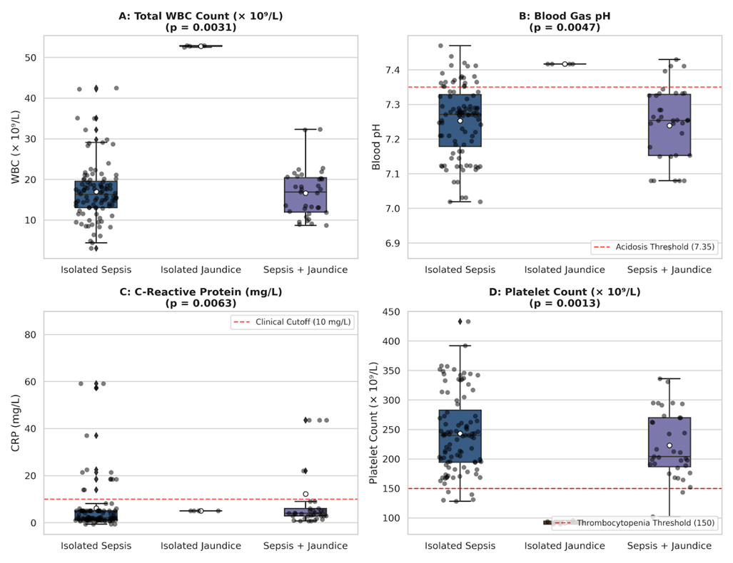
Table (2) provides a comparative analysis of hematological, inflammatory, and metabolic parameters across the three study groups. The most pronounced difference occurred in total WBC counts (P = 0.0031). The isolated jaundice subgroup (n = 4) exhibited an exceptionally high mean automated WBC count of 52.78 ± 0.20 × 10⁹/L. In contrast, the sepsis-only cohort (n = 96) and the combined sepsis-jaundice cohort (n = 35) recorded significantly lower mean counts of 17.01 ± 7.22 × 10⁹/L and 16.63 ± 5.95 × 10⁹/L, respectively. Post-hoc Dunn-Bonferroni pairwise comparisons confirmed that WBC counts in the isolated jaundice group were significantly higher than in both sepsis-containing cohorts (P < 0.005).
Significant metabolic and acid-base derangements were observed across groups. Sepsis cohorts demonstrated acute metabolic acidosis, with mean blood gas pH values dropping to 7.25 ± 0.10 in isolated sepsis and 7.24 ± 0.12 in combined cases, compared to physiological stability (7.42 ± 0.00) in isolated jaundiced neonates (P = 0.0047; post-hoc P < 0.01). Concurrently, serum bicarbonate (HCO3−) levels were significantly depressed in the sepsis cohorts (18.07–18.24 mmol/L) compared to the jaundice subgroup (22.62 ± 0.05 mmol/L, P = 0.0131).
Regarding inflammatory and cellular differential markers, serum CRP levels remained un-elevated at baseline in isolated jaundice (5.00 ± 0.00 mg/L), whereas marked inflammatory spikes were recorded in the combined sepsis-jaundice group (13.15 ± 23.00 mg/L) and isolated sepsis cases (6.46 ± 12.58 mg/L, P = 0.0063). Platelet counts revealed significant differences (P = 0.0013), with jaundiced infants displaying unadjusted mild thrombocytopenia (92.15 ± 3.26 × 10⁹/L) relative to septic infants (243.21 ± 66.97 × 10⁹/L). Absolute lymphocyte counts also differed significantly (P = 0.0202), remaining preserved at 9.41 ± 0.42 × 10⁹/L in isolated jaundice versus 6.07 ± 2.98 × 10⁹/L in isolated sepsis.
Infants with isolated hyperbilirubinemia exhibited leukocytosis (Fig. 1A) despite normal venous blood pH (Fig. 1B) and normal C-reactive protein (CRP) levels (Fig. 1C).
| Parameter | Sepsis only (n = 96) mean ± SD | Jaundice only (n = 4) mean ± SD | Sepsis + Jaundice (n = 35) mean ± SD | Reference Range | P-value |
|---|---|---|---|---|---|
| CRP (mg/L) | 6.46 ± 12.58 | 5.00 ± 0.00 | 13.15 ± 23.00 | < 10.0 | 0.0063* |
| WBC (× 10⁹/L) | 17.01 ± 7.22 | 52.78 ± 0.20 | 16.63 ± 5.95 | 9.0 – 30.0 | 0.0031* |
| Lymphocyte Count (× 10⁹/L) | 6.07 ± 2.98 | 9.41 ± 0.42 | 9.60 ± 16.31 | 2.0 – 11.0 | 0.0202* |
| Lymphocyte (%) | 36.79 ± 14.75 | 18.06 ± 0.68 | 38.72 ± 22.80 | 20.0 – 55.0 | 0.0090* |
| Blood pH | 7.25 ± 0.10 | 7.42 ± 0.00 | 7.24 ± 0.12 | 7.35 – 7.45 | 0.0047* |
| HCO3− (mmol/L) | 18.24 ± 3.38 | 22.62 ± 0.05 | 18.07 ± 3.36 | 17.0 – 24.0 | 0.0131* |
| Platelets (× 10⁹/L) | 243.21 ± 66.97 | 92.15 ± 3.26 | 223.11 ± 56.50 | 150.0 – 450.0 | 0.0013* |
| PO2 (mmHg) | 75.38 ± 56.52 | 164.45 ± 2.52 | 63.95 ± 45.66 | 50.0 – 90.0 | 0.0186* |
| O2 Saturation (%) | 76.41 ± 25.26 | 98.72 ± 0.30 | 70.73 ± 26.60 | 95.0 – 100.0 | 0.0194* |
* Statistically significant at P < 0.05 level (Kruskal-Wallis test).
Clinical outcomes and treatment patterns across the cohorts are summarized in Table (3). Overall neonatal mortality differed by diagnostic group (p = 0.0379), occurring in 19.8% of the sepsis group (n = 19) and 2.9% of the combined sepsis-jaundice group (n = 1). By contrast, no deaths were recorded among infants with isolated jaundice (0%, n = 0).
Hospitalization duration also varied significantly (p = 0.0313). The mean length of stay for isolated jaundice was 3.50 ± 0.58 days, compared with 11.85 ± 11.88 days for isolated sepsis and 9.31 ± 5.55 days for combined cases.
Empirical antibiotic use was high regardless of diagnosis (p = 0.8149), received by 99.0% of neonates with sepsis (n = 95), 100% with combined sepsis-jaundice (n = 35), and 100% with isolated jaundice (n = 4). Birth weight showed no significant variation between groups (p = 0.2155).
| Diagnostic Group | Total (n) | Improved, n (%) | Mortality Rate, n (%) | Antibiotic Use, n (%) | Birth Weight (kg) (mean ± SD) | Length of Stay (days) (mean ± SD) |
|---|---|---|---|---|---|---|
| Sepsis only | 96 | 77 (80.2%) | 19 (19.8%) | 95 (99.0%) | 2.35 ± 0.81 | 11.85 ± 11.88 |
| Jaundice only | 4 | 4 (100.0%) | 0 (0.0%) | 4 (100.0%) | 2.25 ± 0.03 | 3.50 ± 0.58 |
| Comorbid | 35 | 34 (97.1%) | 1 (2.9%) | 35 (100.0%) | 2.68 ± 0.88 | 9.31 ± 5.55 |
| P-value | — | — | 0.0379* (Chi-Square) | 0.8149 (Fisher's Exact) | 0.2155 (Kruskal-Wallis) | 0.0313* (Kruskal-Wallis) |
* Statistically significant at P < 0.05 level.
4. Discussion
This retrospective study evaluated the comparative diagnostic utility of combining C-reactive protein, venous blood pH, and absolute lymphocyte counts to distinguish systemic neonatal sepsis from transient automated leukocytosis among infants admitted to the Neonatal Intensive Care Unit at Al-Jalaa Maternity Hospital. Although marked leukocyte spikes in jaundiced newborns frequently prompted immediate escalation of clinical concern, the findings revealed a pronounced therapeutic paradox: non-septic jaundiced neonates received empirical broad-spectrum antibiotic therapy at rates virtually identical to those with confirmed systemic infection. This widespread antimicrobial exposure was maintained despite a completely benign clinical trajectory in the isolated jaundice group, characterized by full survival and markedly shorter hospital stays compared to septic infants. These observations align with growing international concern that diagnostic over-reliance on isolated hematological anomalies continues to drive routine antimicrobial overuse in neonatal care, particularly where rapid diagnostic markers are evaluated in isolation rather than within a multi-parameter profile.
This defensive prescribing pattern stems largely from clinical anxiety when encountering extreme automated leukocytosis. Standard neonatal management protocols typically mandate immediate antibiotic initiation upon encountering extreme leukocyte spikes due to concern for occult bacteremia or leukemoid reactions secondary to overwhelming infection [23], [24], Patel, et al. [25]. This clinical dilemma is well illustrated in the case reported by Alatassi, et al. [26], where a newborn with a high leukocytosis (159,000/µL) was treated with potent, broad-spectrum antibiotics for suspected sepsis, despite persistently negative CRP levels and completely sterile blood and cerebrospinal fluid cultures. Importantly, even after a prolonged 62-day hospital stay and maximum antibiotic coverage, the researchers acknowledged that the underlying biological mechanism of their patient's leukocytosis remained poorly understood and unexplained. Similar global patterns were documented in a South African NICU, where empirical antibiotic prescription rates reached 95% [27]. These findings underscore the need for structured diagnostic stewardship programs.
In contrast to the automated leukocyte surges, serum C-reactive protein concentrations in the isolated hyperbilirubinemia cohort remained strictly within baseline limits, standing in sharp contrast to the marked inflammatory elevations observed in septic infants. This inflammatory quiescence was accompanied by normal acid-base homeostasis in jaundiced neonates, providing a clear physiological distinction between benign hyperbilirubinemia and systemic infection. Septic neonates, conversely, exhibited pronounced metabolic acidosis, aligning with observations by [4], Arayici, et al. [28], [29], who linked declining blood gas pH to systemic tissue hypoperfusion, anaerobic glycolysis, and microvascular dysfunction during neonatal sepsis.
Crucially, while severe hyperbilirubinemia can cause optical or cell-sizing interferences on automated hematology analyzers leading to spurious white blood cell counts circulating bilirubin does not impair systemic blood gas homeostasis or stimulate hepatic acute-phase protein synthesis. Venous pH and C-reactive protein thereby serve as robust physiological markers that are not confounded by elevated bilirubin levels. Consequently, evaluating blood gas parameters alongside inflammatory kinetics provides robust diagnostic safeguards, allowing clinicians to effectively exclude systemic sepsis and avoid unnecessary antimicrobial intervention in jaundiced newborns.
The absolute quiescence of C-reactive protein in non-septic jaundiced neonates contrasted with marked acute-phase elevations in septic cohorts underscores the utility of this inflammatory reactant as a highly reliable negative predictor. This finding directly aligns with observations by Beltempo, et al. [30] and Hedegaard, et al. [31], who demonstrated that the primary clinical strength of C-reactive protein lies in its high negative predictive value for ruling out occult neonatal infection. Although multi-center international literature reports more dramatic systemic inflammatory surges during neonatal sepsis Fleischmann-Struzek, et al. [32], the moderate baseline elevations documented at Al-Jalaa Hospital likely reflect early empirical blood sampling or regional variations in microbial virulence. Regardless of these magnitude differences, the complete absence of inflammatory kinetic activation in isolated hyperbilirubinemia confirms that unconjugated bilirubin does not stimulate systemic acute-phase responses. Demonstrating an unreactive inflammatory profile alongside intact venous acid-base equilibrium thereby offers compelling, objective evidence to support the prompt de-escalation and safe discontinuation of empirical antimicrobial therapy in clinically stable newborns.
Differential leukocyte dynamics provided further physiological discrimination between true systemic infection and benign hyperbilirubinemia. While septic neonates demonstrated suppressed absolute lymphocyte counts, jaundiced infants maintained preserved lymphocyte levels. In acute bacterial sepsis, systemic inflammatory cascades frequently induce accelerated lymphocyte apoptosis and drive a bone marrow shift toward granulopoiesis, resulting in marked lymphopenia. This pattern was evident in our septic cohort, aligning with observations by Li, et al. [33], who identified neonatal lymphopenia as a key predictor of confirmed bacterial sepsis and adverse clinical outcomes. Conversely, the preservation of normal lymphocyte counts in jaundiced infants, evaluated alongside baseline C-reactive protein and intact venous pH, confirms the absence of an active systemic immune or inflammatory response.
Furthermore, evaluating birth weight indicated that low-birth-weight infants possess limited physiological reserves, rendering them particularly vulnerable to metabolic decompensation during sepsis and contributing to extended hospital stays, consistent with clinical observations by Schellack, et al. [27] and P.G, et al. [14].
This study offers valuable clinical insights into a frequently encountered diagnostic dilemma in neonatal care, highlighting the utility of a simple, multi-parameter profile to prevent unnecessary antimicrobial exposure. Several methodological considerations should be acknowledged. First, although conducted as a single-center retrospective study at Al-Jalaa Maternity Hospital, this institution serves as a major tertiary referral maternity center utilizing standard diagnostic protocols, supporting the regional relevance of these findings. Second, while the isolated jaundice cohort comprised four neonates, this subgroup accounted for every identified real-world instance of extreme automated leukocytosis in non-septic jaundiced infants over an eight-month consecutive sampling period. Third, routine laboratory monitoring relied on standard automated hematology analyzers without automated NRBC correction, reflecting routine daily clinical workflow and highlighting an important target for prospective multi-center studies that incorporate prospective manual differential counts.
These findings carry direct clinical implications for neonatal antibiotic stewardship programs. In resource-limited or high-volume NICU settings, encountering severe leukocyte spikes in jaundiced neonates frequently triggers defensive prescribing and prolonged hospitalizations. Implementing an integrated screening protocol evaluating venous acid-base equilibrium, C-reactive protein, and absolute lymphocyte counts alongside automated hematology metrics provides clinicians with objective criteria to differentiate benign, transient leukocytosis from true systemic infection. Demonstrating an unreactive C-reactive protein profile together with normal venous pH and preserved lymphocytes enables the confident early de-escalation and safe discontinuation of empirical antimicrobial therapy within 24 to 48 hours in clinically stable infants, thereby reducing unnecessary drug exposure, minimizing NICU stay durations, and mitigating the risk of nosocomial complications.
5. Conclusion
The combined profile of C-reactive protein, blood gas pH, and absolute lymphocyte count provides a practical screening aid for distinguishing neonatal sepsis from suspected non-infectious or spurious leukocytosis in jaundiced infants. Its principal advantages are the use of routinely available tests and the potential to support earlier antimicrobial review and shorter unnecessary neonatal intensive care stays. The interpretation is limited by the single-center retrospective design and the very small isolated-jaundice subgroup (n = 4); therefore, the profile should complement, rather than replace, clinical assessment and microbiological testing. Prospective multicenter validation, including manual differential counts and nucleated red blood cell correction, is required before the approach is incorporated into formal antibiotic-stewardship protocols.
6. Acknowledgment
The authors are grateful to the management and NICU staff of Al-Jalaa Maternity Hospital, Libya, for their cooperation and assistance in collecting data.
7. Funding
No specific funding was received for this study.
8. Data Availability
The datasets generated and analyzed during the current study are not publicly available due to institutional privacy policies regarding patient medical records at Al-Jalaa Maternity Hospital. Access to the underlying records is subject to institutional permission and patient confidentiality requirements.
9. Authors' Contributions
Narjes Mabrouk El Osta conceptualized the study, performed the analysis, drafted the manuscript, and revised it. Abdulmonam Elmuzoughi and Mohamed Algazere collected the data, organized the metadata, and managed the spreadsheets. Fathi Alshourif and Ayad Abud facilitated access to the records, verified data accuracy, and critically reviewed the manuscript.
10. Conflict of Interest
The authors declare no conflicts of interest related to this research, authorship, or publication.
11. References
- Parastatidou, S., Tsantes, A. G., Emmanouil, C. C., Konstantinidi, A., Kapetanaki, A., Sokou, R., (2025), Neonatal hemolytic jaundice: Causes, diagnostic approach, and management., Children (Basel), 12(6), 666.
- Begum, N., Afroze, S., (2018), An overview of neonatal unconjugated hyperbilirubinemia and its management., Bangladesh J. Child Health, 42(1), 30-35.
- Milton, R., (2022), Neonatal sepsis and mortality in low-income and middle-income countries from a facility-based birth cohort: an international multisite prospective observational study., Lancet Glob. Health, 10(5), e661-e672.
- Raturi, A., Chandran, S., (2024), Neonatal sepsis: Aetiology, pathophysiology, diagnostic advances and management strategies., Clin. Med. Insights Pediatr., 18, 11795565241281337.
- Yang, J. L., Wang, H. H., (2023), Interpretation of the 2022 American Academy of Pediatrics guidelines for the management of hyperbilirubinemia in newborn infants., Zhongguo Dang Dai Er Ke Za Zhi, 25(1), 11-17.
- Du, L., Ma, X., Shen, X., Bao, Y., Chen, L., Bhutani, V. K., (2021), Neonatal hyperbilirubinemia management: Clinical assessment of bilirubin production., Semin. Perinatol., 45(1), 151351.
- Celik, I. H., Hanna, M., Canpolat, F. E., Mohan, P., (2022), Diagnosis of neonatal sepsis: the past, present and future., Pediatr. Res., 91(2), 337-350.
- Hornik, C. P., (2012), Use of the complete blood cell count in early-onset neonatal sepsis., Pediatr. Infect. Dis. J., 31(8), 799-802.
- Sharma, D., Farahbakhsh, N., Shastri, S., Sharma, P., (2018), Biomarkers for diagnosis of neonatal sepsis: a literature review., J. Matern. Fetal Neonatal Med., 31(12), 1646-1659.
- Parvez, Y., Mathew, A. G., (2014), Hyperleukocytosis in newborn: a diagnosis of concern., Indian J. Hematol. Blood Transfus., 30(Suppl 1), 131-132.
- Kratz, A., Brugnara, C., (2015), Automated hematology analyzers: state of the art., Clin. Lab. Med., 35(1), xiii-xiv.
- Godon, A., Genevieve, F., Marteau-Tessier, A., Zandecki, M., (2012), Automated hematology analysers and spurious counts Part 3. Haemoglobin, red blood cells, cell count and indices, reticulocytes., Ann. Biol. Clin. (Paris), 70(2), 155-168.
- Gandhi, P., Kondekar, S., (2019), A review of the different haematological parameters and biomarkers used for diagnosis of neonatal sepsis., Hematology, 24(1), 45-52.
- P.G, V., M, S., S, S., V, L., Premkumar, L. K., (2025), Assessing neonatal health: The role of blood gas analysis in predicting neonatal outcomes., Asian J. Med. Health, 23(10), 1-8.
- Petel, D. S., (2025), Empiric antibiotic prescribing practices for gram-positive coverage of late-onset sepsis in neonatal intensive care units in North America., Infect. Control Hosp. Epidemiol., 46(1), 102-104.
- Chirico, G., Loda, C., (2011), Laboratory aid to the diagnosis and therapy of infection in the neonate., Pediatr. Rep., 3(1), e1.
- Yilmaz, A., Kaya, N., Gonen, I., Uygur, A., Perk, Y., Vural, M., (2023), Evaluating of neonatal early onset sepsis through lactate and base excess monitoring., Sci. Rep., 13(1), 14837.
- Iqbal, F., Lewis, L. E. S., Siva, N., K E, V., Purkayastha, J., Shenoy, P. A., (2023), Modulation of gut microbiota: An emerging consequence in neonatal sepsis., Clin. Epidemiol. Global Health, 20, 101245.
- Wiertsema, S. P., van Bergenhenegouwen, J., Garssen, J., Knippels, L. M. J., (2021), The interplay between the gut microbiome and the immune system in the context of infectious diseases throughout life and the role of nutrition in optimizing treatment strategies., Nutrients, 13(3), 886.
- Tang, H., Li, W., Xu, Y., Zhou, M., Hamblin, M. R., Wen, X., (2025), Gut microbiota modulation: a key determinant of atopic dermatitis susceptibility in children., Front. Microbiol., 16, 1549895.
- Mehrotra, I., (2021), Characterization of the blood microbiota in children with Celiac disease., Curr. Res. Microb. Sci., 2, 100069.
- Kabeerdoss, J., (2013), Development of the gut microbiota in southern Indian infants from birth to 6 months: a molecular analysis., J. Nutr. Sci., 3, e2.
- Alatassi, E. U., Sukkar, M., Garrada, F. N., (2019), Sepsis-induced hyperleukocytosis in a preterm., Cureus, 11(9), e5594.
- Arayici, S., Şimşek, G. K., Canpolat, F. E., Oncel, M. Y., Uras, N., Oguz, S. S., (2019), Can base excess be used for prediction to early diagnosis of neonatal sepsis in preterm newborns?., Mediter. J. Hematol. Infect. Dis., 11(1), e2019014.
- Wynn, J. L., Wong, H. R., (2010), Pathophysiology and treatment of septic shock in neonates., Clin. Perinatol., 37(2), 439-479.
- Beltempo, M., Viel-Thériault, I., Thibeault, R., Julien, A. S., Piedboeuf, B., (2018), C-reactive protein for late-onset sepsis diagnosis in very low birth weight infants., BMC Pediatr., 18(1), 16.
- Hedegaard, S. S., Wisborg, K., Hvas, A. M., (2015), Diagnostic utility of biomarkers for neonatal sepsis-a systematic review., Infect. Dis. (Lond), 47(3), 117-124.
- Fleischmann-Struzek, C., Goldfarb, D. M., Schlattmann, P., Schlapbach, L. J., Reinhart, K., Kissoon, N., (2018), The global burden of paediatric and neonatal sepsis: a systematic review., Lancet Respir. Med., 6(3), 223-230.
- Uk, N., Hp, S., Vj, R., Js, K., (1985), Hematological indices for early diagnosis of neonatal septicemia., Indian Pediatr., 22, 287-292.
- Sorsa, A., (2018), Diagnostic significance of white blood cell count and C-reactive protein in neonatal sepsis; Asella Referral hospital, South East Ethiopia., Open Microbiol. J., 12, 209-217.
- Patel, S. J., (2009), Antibiotic use in neonatal intensive care units and adherence with Centers for Disease Control and Prevention 12 Step Campaign to Prevent Antimicrobial Resistance., Pediatr. Infect. Dis. J., 28(12), 1047-1051.
- Schellack, N., Gous, A., (2011), Antibiotic prescribing patterns in a neonatal intensive care unit., South African J. Epidemiol. Infect., 26(1), 23-28.
- Benitz, W. E., (2010), Adjunct laboratory tests in the diagnosis of early-onset neonatal sepsis., Clin. Perinatol., 37(2), 421-438.
- Li, X., Wei, Y., Xu, Z., Li, T., Dong, G., Liu, X., Zhu, Z., Yang, J., Yang, J., (2023), Lymphocyte-to-C-reactive protein ratio as an early sepsis biomarker for neonates with suspected sepsis., Mediators Inflamm., 2023, 9077787.